Human iPSC-Derived Cerebral Cortical Neurons (Plated)
iPSC-derived cerebral cortical neurons
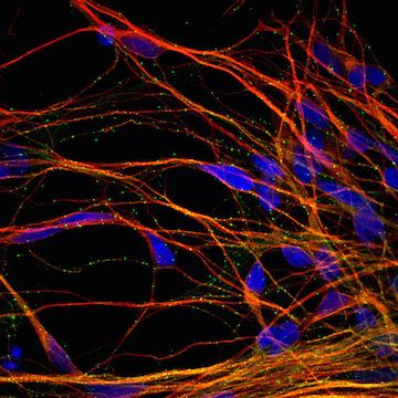
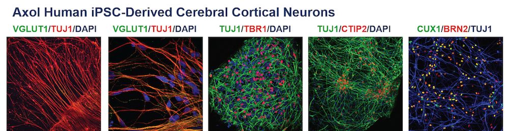
Axol Human iPSC-Derived Cerebral Cortical Neurons express typical markers of cerebral cortical neurons, such as TUJ1, TBR1, CTIP2, BRN2 and CUX1. They are electrically active and can form functional synapses and neuronal networks in vitro.
Axol supplies pre-plated neurons that have been synchronously differentiated from neural stem cells using our fully defined, neuronal differentiation method. Using our specialized coating reagents and Neural Maintenance-XF Medium, the iPSC-derived neurons can be maintained in culture long-term.
Our expert scientists perform the neuronal differentiation and supply neurons in 96-well plate format (center 60 filled). The plated neurons are assay-ready saving you time and resources. The iPSC-derived neurons are shipped live so the neurons are not stressed by cryopreservation. Please inquire for custom plating requests or see the product pages below for further information.
Plated iPSC-derived cerebral cortical neurons from healthy donors
| Cat. No. | Product Name | Starting Material | Quantity |
|---|---|---|---|
| ax0025 | Plated Human iPSC-Derived Cerebral Cortical Neurons (Male) | Cord Blood CD34+ Cells | Center 60 wells of 96-well plate |
| ax0026 | Plated Human iPSC-Derived Cerebral Cortical Neurons (Female) | Cord Blood CD34+ Cells | Center 60 wells of 96-well plate |
| ax0028 | Plated Human iPSC-Derived Cerebral Cortical Neurons (Male) | Fibroblasts (74 Yr Donor) | Center 60 wells of 96-well plate |
| ax0029 | Plated Human iPSC-Derived Cerebral Cortical Neurons (Female) | Fibroblasts (64 Yr Donor) | Center 60 wells of 96-well plate |
Plated iPSC-derived cerebral cortical neurons from patient donors
| Cat. No. | Product Name | Starting Material | Quantity |
|---|---|---|---|
| ax0121 | Plated Human iPSC-Derived Cerebral Cortical Neurons - Alzheimer's Disease Patient (APOE4 HOM) | Fibroblasts (87 Yr Female) | Center 60 wells of 96-well plate (Order here) |
| ax0122 | Plated Human iPSC-Derived Cerebral Cortical Neurons - Alzheimer's Disease Patient (PSEN1 L286V) | Fibroblasts (38 Yr Female) | Center 60 wells of 96-well plate (Order here) |
| ax0123 | Plated Human iPSC-Derived Cerebral Cortical Neurons - Alzheimer's Disease Patient (PSEN1 M146L) | Fibroblasts (53 Yr Male) | Center 60 wells of 96-well plate (Order here) |
| ax0124 | Plated Human iPSC-Derived Cerebral Cortical Neurons - Alzheimer's Disease Patient (PSEN1 A246E) | Fibroblasts (31 Yr Female) | Center 60 wells of 96-well plate (Order here) |
| ax0221 | Plated Human iPSC-Derived Cerebral Cortical Neurons - Huntington's Disease Patient | Fibroblasts (Female) | Center 60 wells of 96-well plate (Order here) |
| ax0421 | Plated Human iPSC-Derived Cerebral Cortical Neurons - Epilepsy Patient | Fibroblasts (5 Mo Female) | Center 60 wells of 96-well plate (Order here) |
| ax1021 | Plated Human iPSC-Derived Cerebral Cortical Neurons - Trisomy X Patient | Fibroblasts (74 Yr Female) | Center 60 wells of 96-well plate (Order here) |
Plated neuron maintenance medium
| Cat. No. | Product Name | Product Description | Quantity |
|---|---|---|---|
| ax0032-500 | Neural Maintenance-XF Medium | Xeno-free medium for maintenance of NSCs and neurons | 500 mL |
Characterization
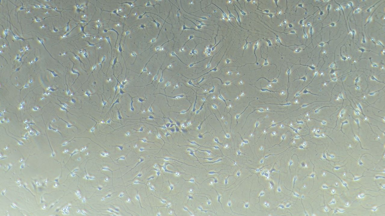
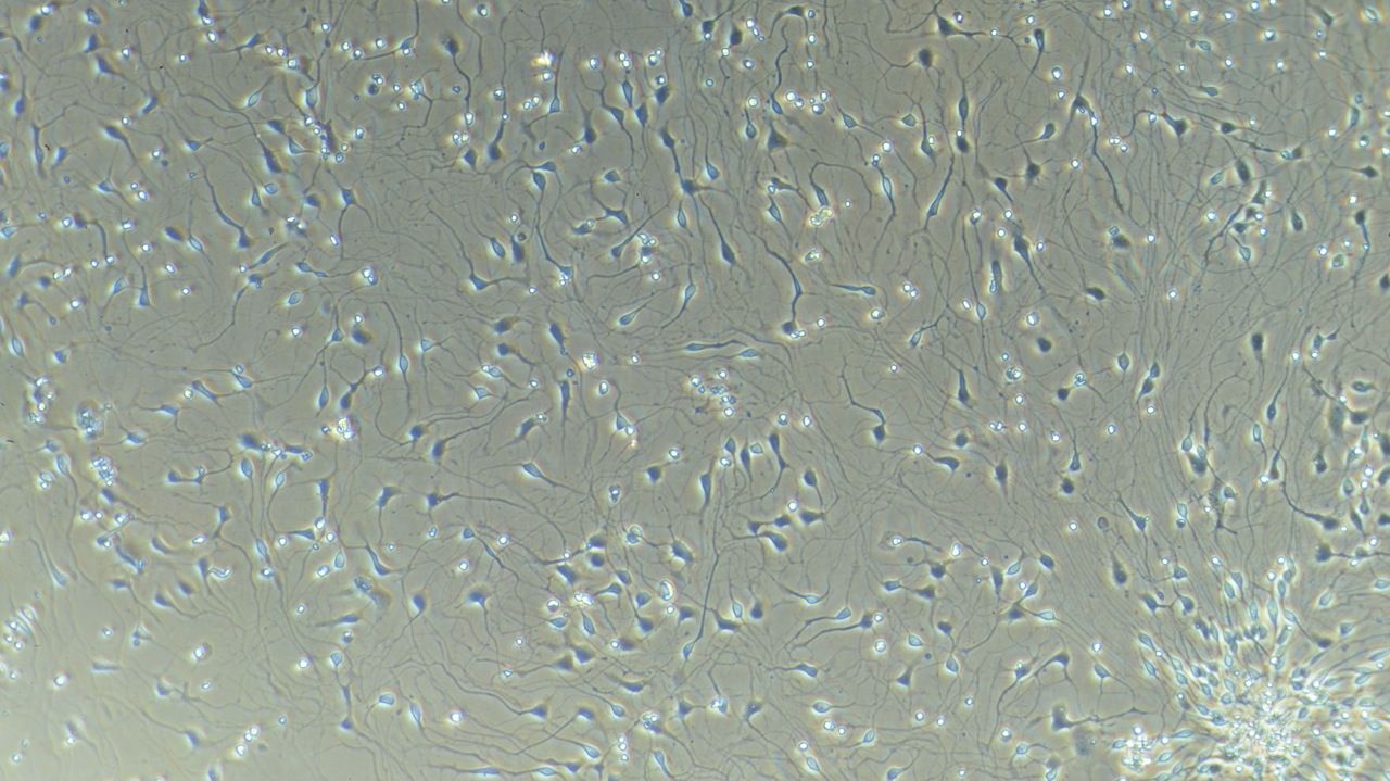
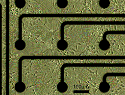
Phase Contrast Images of Axol Human iPSC-Derived Cerebral Cortical Neurons
Immunofluorescence
Transcriptome
Gene expression profiling of Axol cerebral cortical neurons by microarray
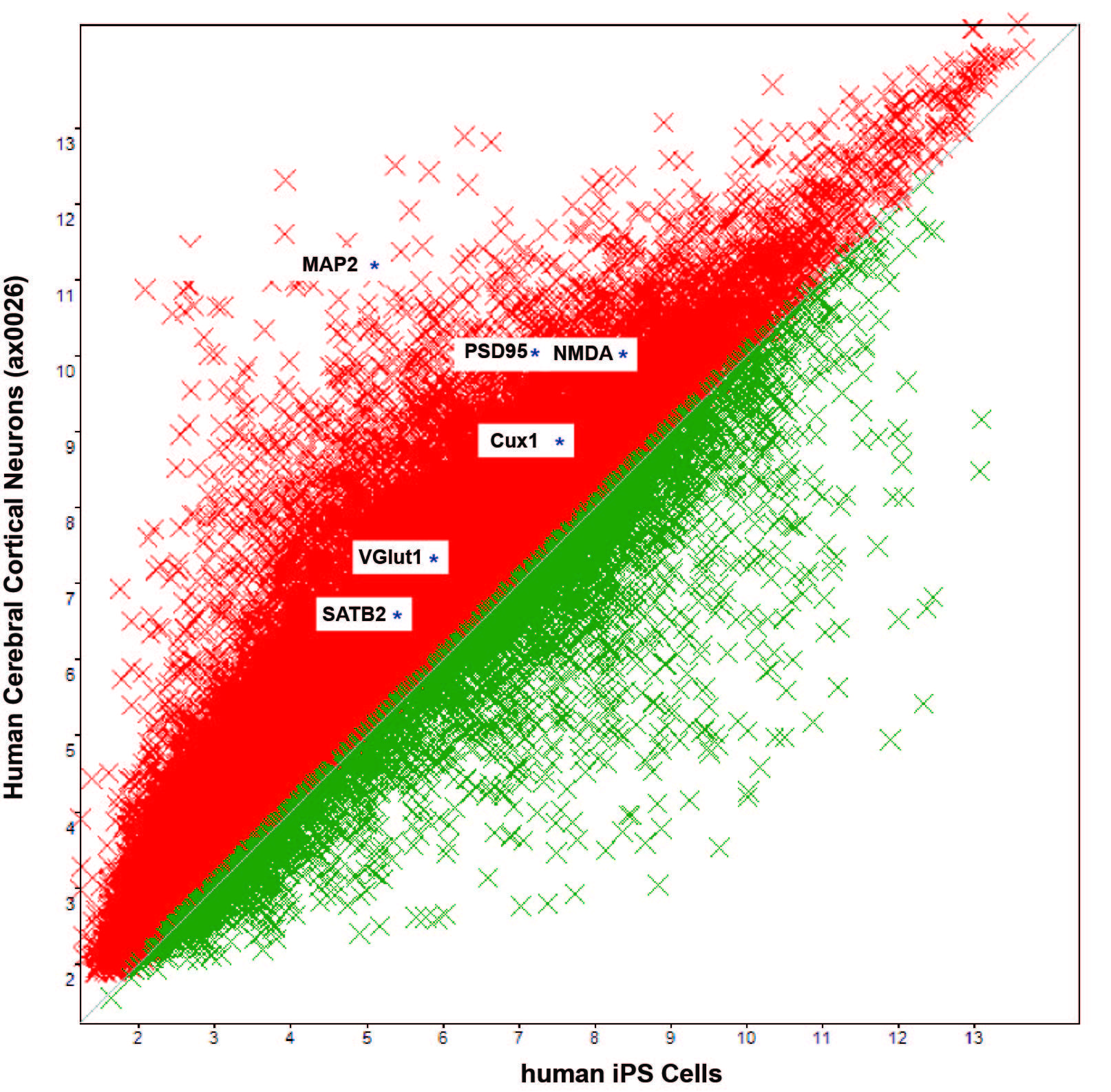
Up-regulated genes in cerebral cortical neurons
 Find the full dataset
The full primary datasets generated for our transcriptome analysis are available at NCBI GEO if you would like to perform your own analysis and look for expression of your gene of interest!
Neurite outgrowth
Live imaging of neurite outgrowth with IncuCyte and NeuroTrackTM by Essen Bioscience Ltd.
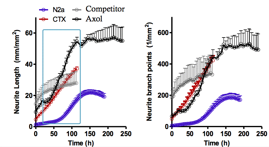
Competitor product comparision
Time-course comparison: N2a mouse neuroblastoma cell line, primary mouse cortical neurons (CTX), competitor's iPSC-derived neurons & Axol's cortical NSC-derived neurons

Live imaging data by Axol Bioscience
Read nowMulti-electrode array (MEA)
MEA analysis of Axol iPSC-Derived Cerebral Cortical Neurons
Human iPSC-derived neurons may be effectively used for drug discovery. Our collaborators, Ikurou Suzuki and his team at Tohoku Institute of Technology in Japan used Alpha MED Scientific’s multi-electrod array (MEA) system to investigate the functional characteristics of human iPSC-derived neurons on their long-term spontaneous activity and drug responsiveness.
The results from this research, using Axol iPSC-Derived Cerebral Cortical Neurons and culture reagents, can be found in Posters and Product Citation sections below.
Drug effects on Axol iPSC-Derived Cerebral Cortical Neurons by Alpha MED Scientific
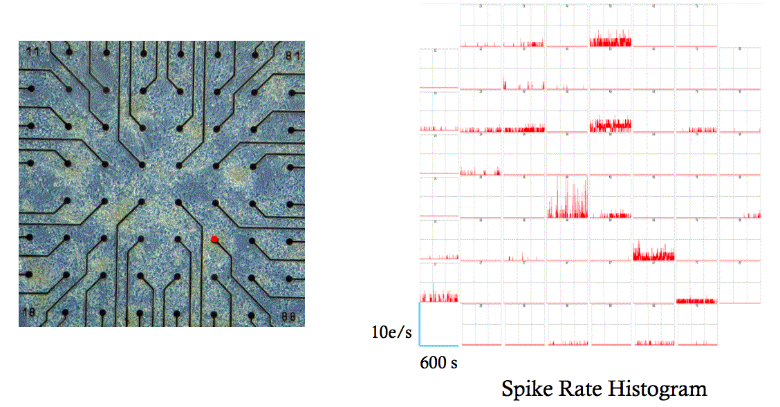
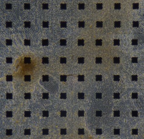 Axol Cerebral Cortical Neurons co-cultured with astrocyte on a MED Probe (MED-P515A, 134 days in vitro)
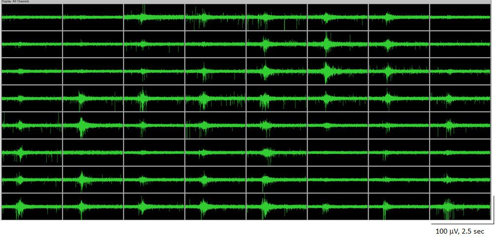
a
64 channel oscilloscope data demonstrating spontaneous activity at 134 Days in vitro
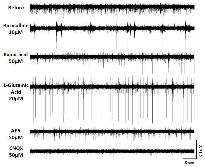
Spontaneous action potentials recorded from Axol Cerebral Cortical Neurons co-cultured with astrocytes (70 days in culture). The traces represent changes in the spontaneous action potentials detected by the same electrodes in response to drug application.
Read more about the performance of Axol cerebral cortical neurons using the MED64 platform
Application of Axol iPSC-Derived Cerebral Cortical Neurons on multi-electrode arrays for studying network electrophysiology by Ole Paulsen's Lab (University of Cambridge)
Spontaneous spiking activity in Axol iPSC-derived neuronal cultures
Complete MEA dataset by Dr Paul Charlesworth
Read nowMEA studies of Axol iPSC-Derived Cerebral Cortical Neurons by Axion Biosystems
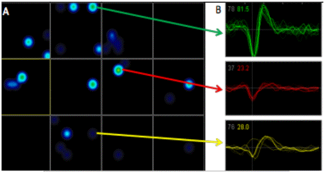
An activity map from the AxIS Software suite showing positions of spike activity for each well of the MEA
iPSC-Derived Cerebral Cortical Neuron morphology on Axion MEAs
Dedicated protocols for using Axol's neural stem cells on Axion MEAs and data generated with Axol's neurons
Read now3D cell culture
3D culture of Axol cerebral cortical neurons in a collagen gel
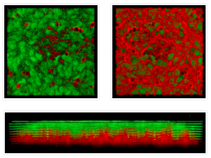
TBR1 / TUJ1
Left Panel: Top View
Top Right Panel: Bottom View
Bottom Panel: Side View
On culturing the cerebral cortical neurons on top of a collagen gel, they formed a uniform static layer of cell bodies. Neurites projected out of these cells and grew downwards, creating a network of interconnected neurites. The results demonstrate that the collagen matrix was suitable for culturing cerebral cortical neurons to achieve neurite outgrowth, although little neuronal migration was observed.