Human iPSC-Derived Microglia

iPSC-derived microglia
Axol’s Human iPSC-derived Microglia provide a reproducible, physiologically-relevant model in an easy-to-use, assay-ready format for investigating neuroglia involvement in neurodegeneration and neurodevelopment. We also offer a fully optimized serum-free Microglia Maintenance Medium to promote and support the maintenance of microglia.
Microglia are commonly described as the immune cells of the brain, with key roles in brain development, neurogenesis, synaptic plasticity and homeostatic maintenance.
Axol’s method for generating iPSC-derived microglia mimics the in vivo pathway of development for brain resident macrophages and produces microglia that are representative of primary human microglia in vitro. Our iPSC-derived Microglia deliver the advantage that they provide an almost infinite source of microglia from a single donor with a normal karyotype. The Microglia are then cryopreserved at > 1 million cells per vial for ease of use.
The homogenous and reproducible population of our iPSC-derived Microglia exhibit physiologically relevant functionality as they are highly phagocytic and produce cytokines in response to pathogens. Our iPSC-derived Microglia also express the microglia-specific marker TMEM119 along with myeloid markers TREM2 and IBA-1. These phenotypes make iPSC-derived Microglia suitable models for investigating neuroinflammation in Alzheimer’s disease, multiple sclerosis and Parkinson’s disease.
iPSC-derived microglia
| Cat. No. | Product Name | Starting Material |
Quantity |
|---|---|---|---|
| ax0666 | Human iPSC-derived Microglia (Ready-to-use live cells) | Cord blood CD34+ cells derived iPSCs | Contact us directly to discuss your specific requirements |
| ax0668 |
Human iPSC-derived Mature Microglia: Cryopreserved | Cord blood CD34+ cells derived iPSCs |
1 cryopreserved vial: > 1 million cells per vial |
| ax0678 | Microglia Complete Cell and Media Kit | Mature Cryopreserved Microglia and Microglia Maintenance Media Kit |
1 vial of ax0668 and 1 media Kit of ax0660 |
Microglia culture media and reagents
| Cat. No. | Product Name | Quantity | |
|---|---|---|---|
| ax0660 | Microglia Maintenance Medium | 100 mL |
Morphological characterization
Brightfield images of Axol’s Human iPSC-Derived Microglia show typical morphology with long ramified processes characteristic of human microglia.

Brightfield images of Axol’s Human iPSC-Derived Microglia.

Brightfield images of Axol’s iPSC-derived Microglia at different stages of maturation. Magnification x20, scale bar = 400um.
Human iPSC-derived microglial precursors (monocytes) were seeded using Microglia Maintenance Medium into 96-well plates at a density of 100,000 cells/cm2. During maturation, spherical monocytes adhere strongly to the culture surface, displaying increasingly ramified morphology as they differentiate to microglia. Axol’s iPSC-Derived Microglia show dynamic surveillance behaviour, which is representative of their phenotype in vivo.
Immunocytochemistry
Human iPSC-derived Microglia show protein expression of myeloid and microglia-specific markers IBA-1, CX3CR1, P2RY12 and TMEM119.

Immunocytochemistry of Axol’s iPSC-derived microglia at Day 18 of in culture.
Immunocytochemistry of Human iPSC-derived Microglia, after 18 days in culture, revealed the expression of IBA-1 and CX3CR1 (a chemokine receptor) as well as the microglial-specific markers TMEM119 (a transmembrane protein), and P2RY12 (purinergic receptor). All 3 markers co-localized strongly with the myeloid marker IBA-1, confirming identity of brain-resident-like microglia.

Axol’s iPSC-derived microglia are a homogenous population. (184/194) x100 =∼95% Pure Population
Differentiation yields microglia that are >95% pure population as assessed by proportion of cells expressing IBA-1 versus P2RY12, in line with similar findings from other groups (Abud et al., 2017; DOI 10.1016/j.neuron.2017.03.042)
Microglia comparison with macrophages
Human iPSC-Microglia express expected myeloid markers and are highly enriched for microglial-specific genes compared to Axol’s iPSC-derived Macrophages derived from the same donor.

A. Microglia and macrophages from the same donor were stained in parallel for the macrophage/microglia markers IBA-1 (voltage-gated Ca2+ sensor) and CX3CR1 (chemokine receptor) as well as microglial-specific markers P2RY12 (purinergic receptor) and TMEM119 (transmembrane protein). Expression of P2RY12 and TMEM119 is either absent or significantly less in macrophages compared to microglia, confirming microglial identity. x10 Magnification; Scale bar=400um
B. mRNA expression of 6 key microglial signature genes in iPSCs, iPSC-derived monocytes, iPSC-derived macrophages and iPSC-derived microglia were determined by qRT-PCR. Cells were lysed from starting iPSCs, Day0 (monocytes), Day14 (macrophages) or Day18 (microglia) prior to RNA isolation followed by RT. CQ1A, MERTK, P2RY12, GPR34, and TMEM119 are all differentially upregulated in microglia. The disease-associated marker TREM2 is expressed at roughly constant levels across the myeloid cell lineage. Fold change was calculated using the ΔΔCT method, with PDGB as an endogenous control and normalisation to iPSC. N=3 (iPSC), N=2 (macrophage), N=1 (monocyte, microglia), technical replicates were performed in triplicate (monocyte, macrophage, microglia) or duplicate (iPSC). Error bars are +- S.E.M.
Axol’s Human iPSC-derived Microglia secrete cytokines in response to inflammatory stimuli

Secretion of the pro-inflammatory cytokine, Tumour Necrosis Factor – alpha (TNF-α), was measured from the cellular supernatants of Axol’s Human iPSC-derived Microglia and Human iPSC-derived Macrophages, using an AlphaLISA. Both microglia and macrophages, from the same donor, showed secretion of TNF-α exhibiting physiological function.

Phagocytosis assay
Phagocytosis was used to measure functional activation and inhibition of Human iPSC-derived Microglia. Phagocytic activity was assessed using pHrodo green Zymosan bioparticles and Human iPSC-derived Microglia were found to be highly phagocytic.

Axol’s iPSC-derived microglia efficiently phagocytose pHrodo green Zymosan particles
A) pHrodo is a fluorogenic dye that dramatically increases in fluorescence as the pH of the surroundings becomes more acidic, such is the case in the lysosome. pHrodo® Green Zymosan BioParticles® (Life Technologies) were added at 0.5 mg/mL to day 19-microglia plated at 1x10⁵sup>/cm2, and incubated for 3hrs at 37oC, 5% CO2. Nuclei were counterstained with a live Hoechst 33342 stain and live cell imaging was carried out using an on-stage incubator connected to an EVOS Fl Auto imaging system (Life Technologies). Two activated microglial cells can be seen consuming increasing numbers of BioParticles® over time leading greater and more intense fluorescence emission. B) and C) Microglia seeded at 1x10⁵/cm2 (B) or 3x105cm2 (C) were pre-treated with 100 ng/mL lipopolysaccharide (LPS) for 1hr (B), 5, 10μM or 30μM Cytochalasin D (CytoD, phagocytosis inhibitor) or 1μM or 10μM Latrunculin A (Lat-A) for 30mins (B) or 1hr (C) before adding pHrodo® Green Zymosan BioParticles® at 0.5 mg/mL and incubating for 3hrs at 37oC, 5% CO2. Lat-A inhibited phagocytosis more strongly than CytoD, in line with Lat-A being a more potent inhibitor of actin polymerization. Fluorescence was quantified using a SpectraMax iD3 microplate reader (Molecular Devices), and values were blanked against a no-cell control well. N=4 for both experiments, One-way ANOVA *P<0.05, **P<0.01, ***P<0.001
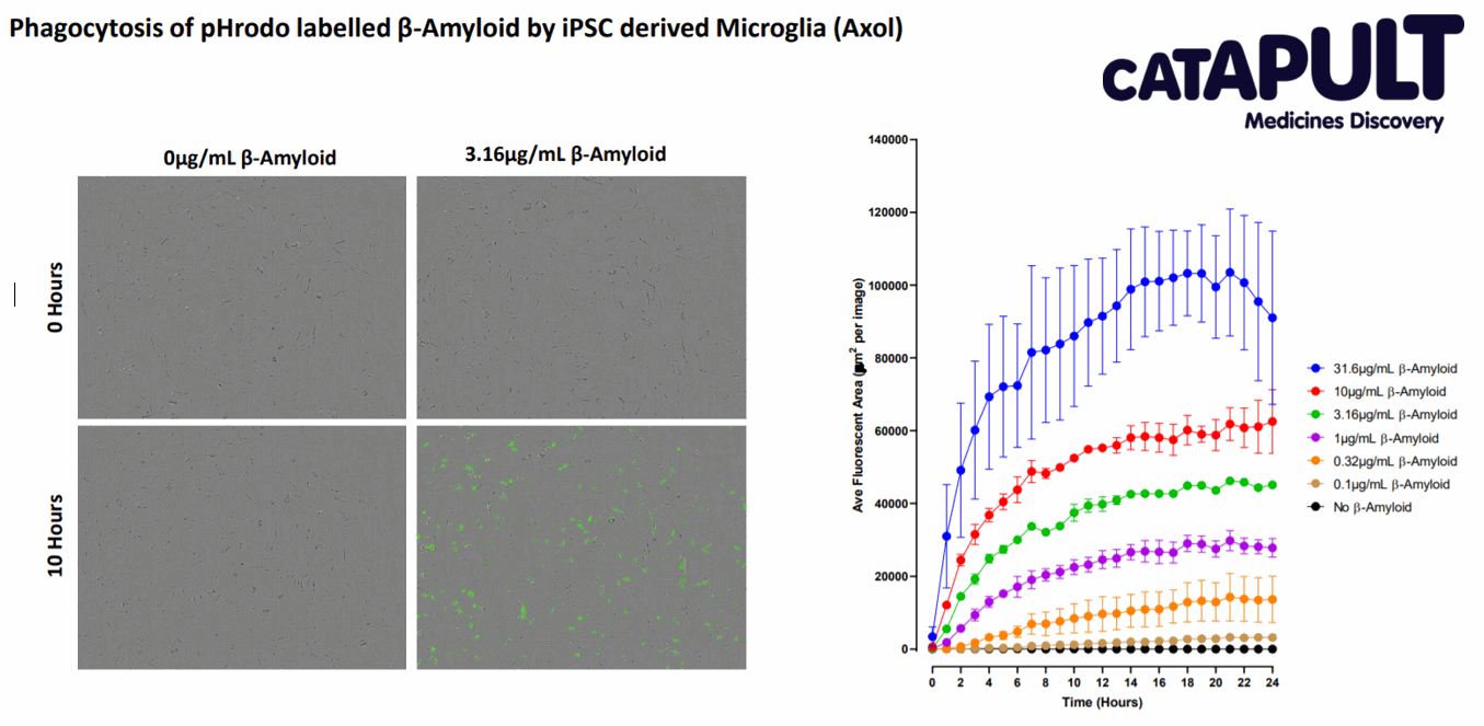
Further Functional Phagocytosis data (courtesy of Medicines Discovery Catapult; Alderley Edge, Cheshire, UK)
Phagocytosis is used as a primary measure of functional Microglia activity. The Cryopreserved Microglia (ax0668) exhibit substrate-dependent phagocytosis when exposed to fluorescent pHrodo labelled beta-amyloid. The Human IPSC derived cryopreserved microglia are shown to be highly phagocytic in nature and equivalent to fresh cells.
Cytokine response
Cytokine Responses in iPSC-microglia

A and B) Stimulation with TLR agonists LPS (TLR1/2) and R848 (TLR7/8), as well as the cytokine IL1β and interferon gamma (IFNγ) induces changes in TNFα and IL-6 release.
Microglia plated at 120,000/cm2 were stimulated with increasing concentrations of LPS, R848, IL1β, and IFNγ for 20hrs. The concentration of TNFα (A) and IL-6 (B) in the harvested supernatant were measured by alphaLISA. Except for LPS, there is a clear dose-dependent, positive correlation between simulant concentration and amount of cytokine released. N=1. Data kindly provided by Grünenthal GmbH, Germany.
Co-culture of microglia and cerebral cortical neurons
Axol’s Human iPSC-derived Microglia are suitable for co-culture with Human iPSC-derived Cortical Neurons.

A. Immunocytochemistry of Human iPSC-derived Microglia co-cultured with Human iPSC-derived cortical neurons reveals microglia specific maker expression TMEM119 and neuronal marker β-3 Tubulin.

B. Phagocytosis analysis revealed activation of microglia after addition of pHrodo green zymosan particles (0.15 mg/mL) and inhibition after treatment with CytoD (10 μM) however no further stimulation was observed when stimulated with LPS (100 ng/mL).

C. Images taken during phagocytosis assay showing the effect of LPS-induced clustering can be seen with several microglia in close proximity responding to and engulfing the pHrodo® Green Zymosan BioParticles®.

D. Cytokine response was analysed from Human iPSC-derived Microglia co-cultured with Human iPSC-derived Cortical Neurons from the same donor, after stimuli with increasing concentrations of LPS. The co-culture were incubated with LPS for 5 hours at 37oC . Bars represent mean ±S.E.M, N=3 independent experiments. TNF-α ELISA revealed an increasing secretion of cytokine, TNF-αα with increasing LPS concentration.
Download the co-culture application protocolCo-culture of the Cortical Neurons, Astrocytes and Microglia

A co-culture model of neurons, astrocytes and microglial cells were infected with the IncuCyte® NeuroBurst Orange Reagent (a genetically-encoded fluorescent calcium sensor). Calcium-flux kinetics were captured and analysed. Data kindly provided by Sartorius.
Resources
Protocol
Application Note
- Characterization of human iPSC-derived Microglia using live-cell imaging reveals hallmarks of morphology and function
-
This study characterized human iPSC-derived microglia using IncuCyte® Live Cell assays (Essen BioScience, part of the Sartorius Group) to validate their morphological and functional features, offering insights into their suitability for immune and neurodegenerative disease research. Live-cell functional assays quantified the ability of human iPSC-derived microglia to phagocytose apoptotic neuronal cells (efferocytosis) and neurodegenerative disease associated peptides.
Frequently Asked Questions for Human iPSC-derived Microglial Precursors (ax1666)
Do these cells come in frozen vials?
No, these cells are shipped live in cryovials and must be plated immediately upon receipt.
What is the purity of the microglia culture?
Our microglia are ~95% pure, as assessed immunocytochemically by co-localisation of IBA-1 (myeloid marker) and P2RY12 (microglia-specific marker).
How do you ensure lot-to-lot consistency and what are the QC criteria?
Microglia Precursors (ax1666) and required Medium Kit (ax0660) are tested for sterility and mycoplasma contamination. Cells must be >90% viable pre-shipment and test positive for the presence of IBA-1, CX3CR1, TMEM119 and P2RY12 after a 2-week differentiation to be considered as QC-passed.
Is it possible to expand microglial precursors?
No, the precursors are non-proliferative and will begin to differentiate upon attachment to cell-culture treated surfaces
How long can the microglia be kept in good condition after 2 weeks of differentiation?
A maximum of 1 week. After this, cells will begin to die off, which may affect downstream assays.
How often are microglial precursors produced? Is it possible to specify a shipping date?
The cells are made on a continuous rolling production, so should be readily available to purchase year-round. Each lot of cells produces for 4 weeks and vials can be reserved for shipment on a weekly basis over this period. Availability therefore depends on the number of reservations already placed for certain weeks of a given lot.
Is the differentiation/maturation media provided?
Accompanying media is sold separately as Microglia Maintenance Medium (ax0660), and its use specifies differentiation/maturation for 2 weeks and subsequent maintenance of microglia for an additional 1 week maximum if desired.
Are these cells similar to HSCs
These cells are quite different from blood-derived monocytes, in terms of ontogeny and gene expression profile. The generation of our iPSC-microglial precursors mimics the in vivo, myb-independent pathway of differentiation. Our cells are not HSC or HSC-like as these are by definition dependent on MYB for their self-renewal. That said, they express some classical BDM markers, being CD45+/CD11b+/CD14+ and can be differentiated to iPSC-macrophages (https://www.axolbio.com/page/ipsc-derived-macrophages) as well as iPSC-microglia. Differentiation to other lineages may be possible though it is at the purchaser’s own risk.
Can the microglial precursors be detached and re-seeded?
Once plated, the microglial precursors adhere very strongly to cell culture-treated surfaces and are very difficult to detach without use of a cell scraper, which has the potential to cause mechanical damage and/or activate the cells. Passaging using Accutase with gentle scraping has been tested, and while some cells do reattach, the ramified morphology is lost and does not recover.As such, we strongly recommend plating into final format.
Can the cells be seeded onto glass and if so, is there a recommended coating?
Microglial precursors attach readily to glass slides/coverslips/wells and can be seeded directly onto untreated glass surfaces. Differentiation can be carried out for 2 weeks in maintenance medium as normal
How long does shipping take on average?
Usually 1-2 days depending on geographic location. In some circumstances, it may take up to 3 days. We have successfully tested a 3-day shipment of live microglial precursors with no detrimental effects to the cells or differentiation capacity.
Can the cells be polarised to M1 or M2 phenotypes?
Yes, the cells can be polarised to M1 or M2 phenotype using appropriate cytokine stimulation, and adopt the functions of that phenotype.
Frequently Asked Questions for Human iPSC-derived Microglia (ax0666)
What is the purity of the microglia culture?
Our microglia are ~95% pure, as assessed immunocytochemically by co-localisation of IBA-1 (myeloid marker) and P2RY12 (microglia-specific marker).
How long can the microglia be kept in good condition?
A maximum of 1 week. After this, cells will begin to die off, which may affect downstream assays.
Is the differentiation/maturation media provided?
Accompanying media is sold separately as Microglia Maintenance Medium (ax0660), and its use specifies maintenance of microglia for 1 week maximum.
How long does shipping take on average?
Usually 1-2 days depending on geographic location. In some circumstances, it may take up to 3 days.
Can the cells be polarised to M1 or M2 phenotypes?
Yes, the cells can be polarised to M1 or M2 phenotype using appropriate cytokine stimulation, and adopt the functions of that phenotype.