Human iPSC-Derived Renal Proximal Tubular Cells
Renal Proximal Tubular Cells
Axol iPSC-Derived Renal Proximal Tubular Cells are derived from integration-free iPSCs of a healthy male donor and have been differentiated through the up-regulation of specific signalling pathways. To support the growth and maintenance of Axol iPSC-Derived Renal Proximal Tubular Cells, we offer our Renal Epithelial Cell Culture Medium which is fully supplemented with hormones and recombinant growth factors that promote optimal growth.
Our iPSC-Derived Renal Proximal Tubular Cells express critical functional markers, along with epithelial and renal cell markers. These include the important phosphate transporter, SLC34A1, the organic ion transporter crucial for baso-lateral uptake of organic anions, OAT1 and the apical glucose transporter SGLT2, key in glucose reabsorption from the glomerular filtrate. These cells provide a physiologically relevant model for investigating glucose uptake in diabetes, the transportation of drugs and endogenous metabolites, and detecting nephrotoxicity. Axol iPSC-Derived Renal Proximal Tubular Cells are ready for endpoint assays within 4 days of plating providing an easy-to-use, assay-ready system for cell based assays.
Human iPSC-derived renal proximal tubular cells
| Cat. No. | Product Name | Starting Material | Quantity |
|---|---|---|---|
| ax2115 | Human iPSC-derived Renal Proximal Tubular Cells (Male) | Cord Blood CD34+ Cells | ≥1 million cells |
Renal epithelial cell culture media
| Cat. No. | Product Name | Quantity |
|---|---|---|
| ax3534 | Renal Epithelial Cell Culture Media | See Product Page |
Phase contrast
Phase contrast images show the characteristic morphology of Axol Human iPSC-Derived Renal Proximal Tubular Cells.

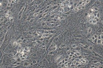
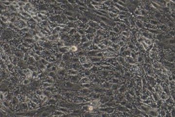
Phase contrast images of Axol Human iPSC-Derived Renal Proximal Tubular Cells at day 4 post-thaw. Human iPSC-derived Renal Proximal Tubular Cells are seeded using Renal Epithelial Cell Culture Medium at a density of 50,000 cells/cm2. The iPSC-derived renal proximal tubular cells form an adherent, confluent monolayer with typical epithelial morphology by day 4 after seeding.
Immunocytochemistry
Axol iPSC-Derived Renal Proximal Tubular Cells show the expression of key proximal tubular cell markers in the correct subcellular localization.
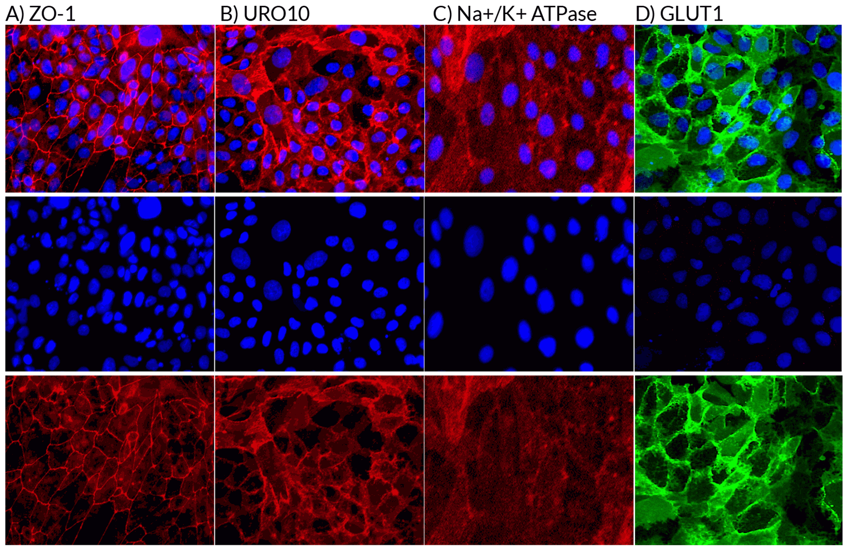
Expression of the ZO-1, URO10, Na+/K+ ATPase and GLUT1 proteins were confirmed in Axol iPSC-Derived Renal Proximal Tubular Cells using immunocytochemistry (A, B, C and D). Staining for ZO-1, URO10 and Na+/K+ ATPase (red) and GLUT1 (green) respectively confirmed the presence of tight junctions (A), urothelial glycoprotein (present on the surface of proximal tubule cells) (B), sodium-potassium pumps (C) and the uniporter protein, glucose transporter 1 (D). DAPI counterstain.
Gene Expression
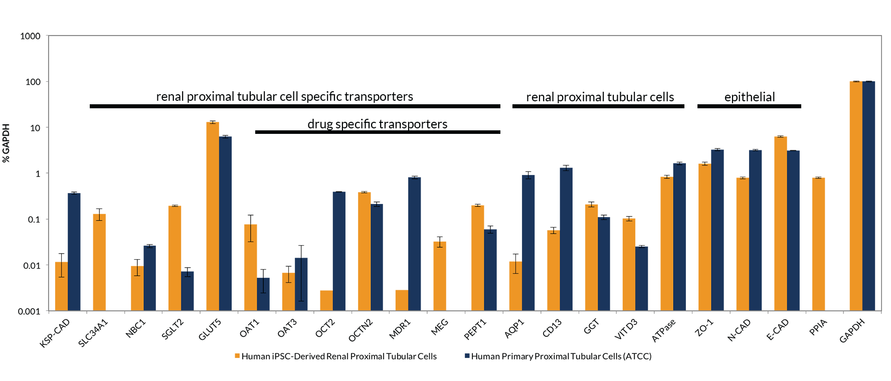
Quantitative real-time reverse transcription polymerase chain reaction (RT-qPCR) analysis was used to determine the gene expression levels of 22 genes that identify renal proximal tubular cells.
RT-qPCR reveals the expression of 22 genes important for proximal tubular cell function. Key transporter genes that are not highly expressed in human primary renal proximal tubular cells such as SLC34A1 and SGLT2 are expressed in iPSC-Derived Renal Proximal Tubular Cells. The expression of these genes supports the use of Axol iPSC-Derived Renal Proximal Tubular Cells in transportation assays and in the detection and prediction of nephrotoxicity.
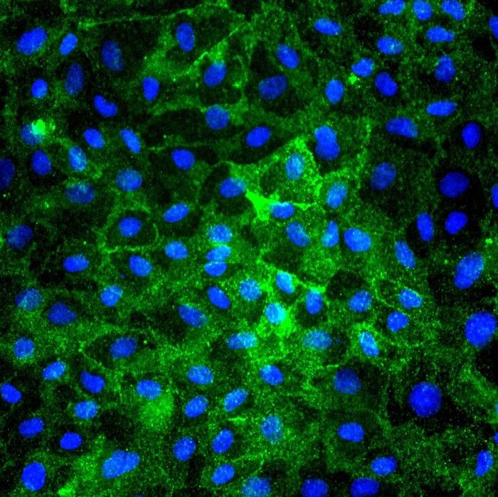
Albumin Endocytosis
Albumin endocytosis in iPSC-Derived Renal Proximal Tubular Cells
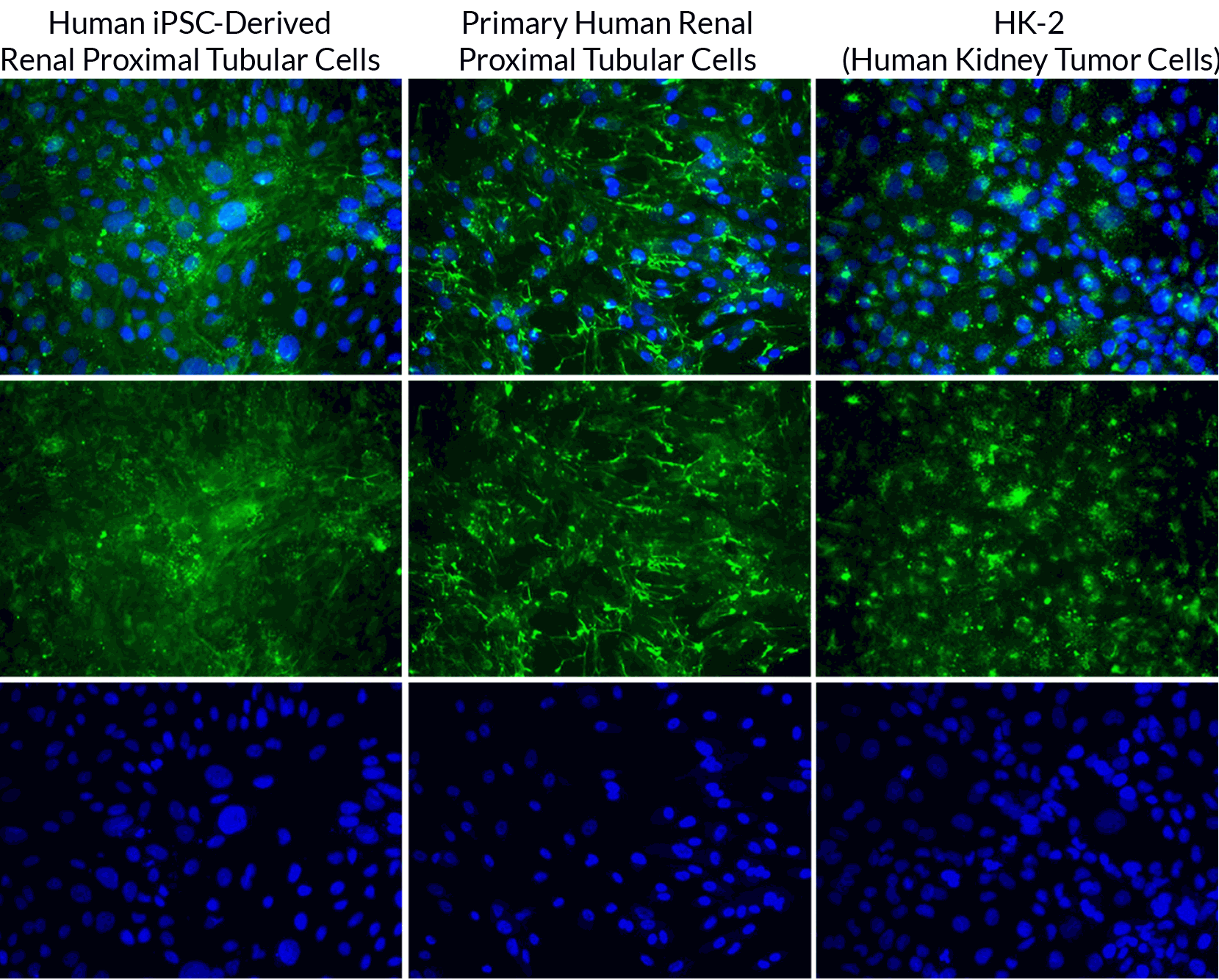
Human iPSC-Derived Renal Proximal Tubular Cells show albumin reabsorption.
iPSC-derived renal proximal tubular cells, primary human renal proximal tubular cells and human kidney tumor cells were treated with 100 µg/mL FITC-conjugated BSA in serum-free DMEM-F12 for 16 hours, in parallel. The FITC-BSA can be seen in the cytoplasm of each cell type signifying that the renal proximal tubular cells are able to endocytose albumin.